Translational Cancer Scientist · UCLA David Geffen School of Medicine

Advancing the science of solid tumor therapeutics.

Senior translational scientist bridging in-vivo oncology, pancreatic cancer biology, and preclinical drug development — from mechanism to medicine.

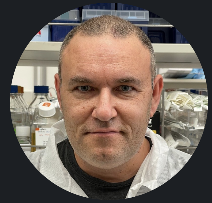
Portrait of Yaroslav Teper, PhD, in the laboratory
Pancreatic Cancer Translational Lab
15+
Years in translational oncology
10+
Peer-reviewed publications
2
First-in-class compounds advanced
4
Leading research institutions
About

Turning pancreatic tumor biology into preclinical therapeutics.

I lead translational research at the intersection of metabolism, immunology, and cancer — designing the in-vivo models and screening programs that move promising molecules toward the clinic.

I'm a senior scientist with deep expertise in in-vivo drug screening, solid tumor oncology, and preclinical efficacy studies. My career has centered on the hardest questions in pancreatic cancer — how obesity, chronic stress, and inflammation converge to drive tumor initiation and progression.

As co-head of the Pancreatic Cancer Translational Laboratory at UCLA, I combine sophisticated murine models with computational biology and digital pathology to identify actionable targets and repurpose approved drugs as chemo-preventive agents. Earlier, I played pivotal roles developing the anti-metastatic compound Metarrestin and the immunomodulatory peptide RP-182.

Pancreatic cancer (PDAC) Tumor microenvironment In-vivo efficacy Cancer metabolism Tumor immunology Drug repurposing

In-Vivo & Laboratory Methods

Complex murine model generation (inducible KrasG12D), high-throughput phenotypic screening, target deconvolution, flow cytometry, and tumor microenvironment modulation.

Bioinformatics & Programming

Python and R for genomic/transcriptomic analysis of FASTQ sequencing data, statistical computing, experimental modeling, and data visualization — accelerated with cloud and AI tooling.

Digital Pathology & Imaging

Advanced QuPath workflows for quantitative digital pathology, automated IHC quantification, and biomarker evaluation (e.g., GLP1R).

Research Focus

Three questions driving my work.

My research program spans the biology of pancreatic cancer, the development of first-in-class therapeutics, and the computational tools that make discovery faster.

01

Metabolism, stress & PDAC

Investigating how diet-induced obesity and chronic stress converge on CREB phosphorylation to accelerate pancreatic cancer — and how approved drugs like metformin, statins, and beta-blockers might interrupt early carcinogenesis.

KrasG12D modelsChemo-preventionCREB signaling
02

First-in-class therapeutics

Preclinical development, screening, and target deconvolution of novel oncology compounds — including the anti-metastatic agent Metarrestin (ML-246) and the CD206-targeting host-defense peptide RP-182.

MetarrestinRP-182In-vivo efficacy
03

Tumor immunology

Characterizing macrophage-driven acinar-to-ductal metaplasia and inflammatory cytokine production to uncover immunotherapeutic vulnerabilities and reshape the pancreatic tumor microenvironment.

MacrophagesADMImmunotherapy
Research Spotlight

From target discovery to first-in-class therapeutics.

Four studies that trace my work across the drug-discovery pipeline — from the genomic discovery and validation of a novel pancreatic cancer target, to the signaling mechanisms that drive the disease, to the preclinical development of a first-in-class anti-metastatic drug, to the computational biology that mines single-cell data for new immune targets.

1Developing Metarrestin — a first-in-class anti-metastatic drug

Metarrestin (ML-246) is a first-in-class small molecule that disrupts the perinucleolar compartment — a structure found almost exclusively in metastatic cancer cells — and inhibits RNA polymerase I transcription. In preclinical models it suppressed metastasis across three cancer types while remaining remarkably well tolerated, and it has since advanced toward first-in-human clinical studies. The work was published in Science Translational Medicine (2018).

My contribution

I developed the NSG / PANC1 metastatic pancreatic cancer xenograft model and conducted the initial in-vivo efficacy studies that established proof of concept — demonstrating that metarrestin could halt metastatic progression and extend survival in a living system.

Dosed at 10 mg/kg through the diet, metarrestin-treated mice bearing metastatic PANC1 tumors had no mortality beyond 90 days, while control mice began dying at day 25. Treatment more than doubled median survival in advanced-disease animals, significantly reduced metastatic burden in both liver (p < 0.01) and lung (p < 0.05), and preserved organ architecture — all with no measurable impact on body weight or behavior.

>90d
No mortality on treatment (vs. day 25 for controls)
2×+
Median survival extension in advanced disease
p<0.01
Reduced liver metastatic burden

In-vivo efficacy — PANC1 metastatic xenograft

Survival after start of treatment (early / micrometastatic cohort)

100 75 50 25 0 0 25 50 75 90+ Days after start of treatment Survival (%) Metarrestin Control
Schematic of the reported survival result (Fig. 3A) — redrawn from the published data, not the original figure. Metarrestin (10 mg/kg in diet) vs. control in the PANC1 metastatic xenograft model. Source: Frankowski, Wang, Patnaik, … Teper Y, et al. "Metarrestin, a perinucleolar compartment inhibitor, effectively suppresses metastasis." Sci. Transl. Med. 2018. Read the paper →

2Target discovery & validation — Plexin A1 in pancreatic cancer

To uncover new drivers of pancreatic cancer, we established three patient-derived pancreatic cancer cell lines and whole-genome-sequenced two of them against matched normal DNA. Among the somatic variants, we pinpointed a previously unreported mutation in PLXNA1 — the axon-guidance receptor Plexin A1 — c.2587G>A (D863N), a gene family already tied to poorer pancreatic cancer survival.

My expertise

This study showcases my approach to target discovery and validation — building patient-derived tumor models, mining whole-genome sequencing for candidate drivers, and applying gain- and loss-of-function assays to prove a variant is functional, not incidental.

Overexpressing the mutant receptor drove ligand (SEMA3A)-induced invasion that wild-type PLXNA1 did not, while silencing it cut invasion by ~50% and selectively reduced proliferation in the mutant-bearing line. Together, these gain- and loss-of-function results identified the single base change as an oncogenic gain-of-function driver — and nominated axon-guidance signaling as a therapeutic target in pancreatic cancer.

3
Patient-derived lines established & sequenced
D863N
Novel PLXNA1 gain-of-function variant
~50%
Drop in invasion on knockdown

Target discovery & validation pipeline

From patient tumor to a validated driver mutation

1

3 patient-derived cell lines established from pancreatic tumors

2

Whole-genome sequencing of two lines vs. matched normal DNA

3

Novel somatic variant: PLXNA1 c.2587G>A (D863N)

4

Functional validation — overexpression + siRNA knockdown

Gain-of-function driver — ↑ invasion & ↑ proliferation → candidate target

Discovery-to-validation workflow from the study. Source: Sorber R, Teper Y, Abisoye-Ogunniyan A, et al. "Whole-genome sequencing of newly established pancreatic cancer lines identifies novel somatic mutation (c.2587G>A) in axon-guidance receptor Plexin A1 as enhancer of proliferation and invasion." PLOS ONE 2016. Read the paper →

3Mechanism — how stress & obesity converge to drive pancreatic cancer

Chronic stress and diet-induced obesity are both epidemiologically linked to pancreatic cancer — but through what mechanism? We showed the two exposures funnel into the same molecular switch. Stress signals act through the β-adrenergic receptor and PKA; obesity-related neurotensin acts through protein kinase D (PKD); and both converge on phosphorylation of the transcription factor CREB to drive PDAC cell proliferation.

My contribution

As co-first author, I designed and led the in-vivo work — the diet-induced obesity and chronic-stress mouse models that translated this signaling convergence into accelerated pancreatic carcinogenesis.

In Kras-driven mice, a high-fat diet alone raised advanced PanIN-3 precancerous lesions nearly ten-fold (6.1% vs. 0.6%), and adding chronic social-isolation stress pushed them higher still (16.1%) — an effect most pronounced in females, who express more β-adrenergic receptors. Because the stress arm runs through the β-adrenergic receptor, the study points to widely used beta-blockers as a potential chemo-preventive strategy.

2→1
Stress & obesity pathways converge on CREB
16%
PanIN-3 lesions with diet + stress (vs. 0.6% control)
Rx
Beta-blockers as a chemo-preventive lead

Converging signaling — stress, obesity & CREB

Two risk factors, two pathways, one molecular switch

Chronic stress Diet-induced obesity β-adrenergic → PKA Neurotensin → PKD CREB phosphorylation Pancreatic cancer
Signaling convergence described in the study — chronic stress (β-adrenergic → PKA) and diet-induced obesity (neurotensin → PKD) both drive CREB phosphorylation. Source: Sun X, Teper Y, et al. "Stress and Obesity Signaling Converge on CREB Phosphorylation to Promote Pancreatic Cancer." Mol. Cancer Res. 2025. Read the paper →

4Computational biology — mining single-cell data for immune targets

Alongside the bench work, I run the computational side: bulk RNA-seq and single-cell analyses spanning human pancreatic cancer atlases (208,000+ cells across 38 donors) and our own KC mouse cohorts — diet, chronic stress, and beta-blocker arms — to find immune states worth targeting.

My approach

I build the tooling and, just as importantly, the controls that decide what survives — depth matching, sex matching, contamination ceilings, and negative controls. When a public atlas proved too large to load conventionally, I wrote a streaming reader that maps a 3.8 GB single-cell object in about 30 seconds using under 1 GB of memory.

Two findings survived every control. In human PDAC, a fusogenic macrophage population — enriched for the cell-fusion genes DCSTAMP and ATP6V0D2 — that isn’t explained simply by monocyte influx. And in mouse macrophages, CD36⁺ cells show suppressed type-I interferon signaling, reproduced within a single library, so no batch, sex, or dissociation difference can account for it. Several headline results did not survive their controls, and were retired rather than reported.

320k+
Cells analyzed across human & mouse datasets
2
Findings that survived every control
~30s
To map a 3.8 GB atlas with a custom reader

Analysis pipeline & the controls that gate it

From raw sequencing objects to claims that hold up

1

Acquire & stream — public atlases plus our own bulk and single-cell runs; custom reader for oversized objects

2

Process — QC, normalization, clustering, and per-cell annotation rather than per-cluster

3

Score immune programs — macrophage ontogeny, lipid handling, interferon, cell fusion

4

Apply controls — depth & sex matching, contamination ceilings, reciprocity, batch floors

Keep what survives — fusogenic TAMs and CD36→IFN suppression as candidate immune targets

Workflow applied across human PDAC atlases and KC mouse cohorts. Analyses ongoing; the CD36–interferon relationship independently recovers a mechanism reported in Cancer Research (2025). Tooling: Python, scanpy, and a custom streaming reader for R serialization objects.

Processing 70 GB of sequencing data on a 7 GB machine

Transcriptomics files are enormous, and the conventional route is to unzip everything to disk and load it into memory — which effectively requires a dedicated server. Instead I stream: the compressed archives are opened but never unzipped, reads are pulled out a slice at a time under a hard memory ceiling, and each sample is quantified independently so nothing is ever pooled before the count matrix exists.

The payoff is that the peak memory stays flat no matter how large the input, single-cell steps run on sparse matrices with no dense copies, and the final statistics ship as a single self-contained HTML file that still recomputes every comparison in the browser.

Streaming genomics — ~70 GB of sequencing data through a 7 GB machine Nothing decompressed to disk · nothing pooled before the count matrix · statistics delivered as 2.5 MB of self-contained HTML ARCHIVES ON DISK two zip archives 44.2 GB + 25.8 GB opened, never unzipped members streamed out one at a time 70 GB source material STREAM + REPAIR unzip -p head -c N trim both mates to min(R1,R2) cut on a multiple of 4 lines ~120 MB per pull, hard ceiling QUANTIFY selective alignment to a cDNA index one sample at a time, independently 26 × ~4.5M pairs 79.8–92.0% mapped 1.4–1.7 GB peak resident memory ANALYSE differential expression + gene-set enrichment single-cell on sparse matrices, no dense copy 24,013 cells across four libraries 7 GB · 2 cores the entire machine DELIVER self-contained HTML, no server, works offline quantised typed arrays, base64-packed PCA, Fisher, Spearman run in the browser 2.5 MB still fully interactive new experimental arms append — nothing already processed is reprocessed ≈ 28,000× reduction from raw archives to interactive deliverable, with every underlying comparison still recomputable on click.
  • Flat memory, any input sizePeak usage is set by the slice size, not the archive — so the same pipeline runs on a laptop.
  • Nothing touches diskArchives are read in place. No decompressed intermediates to write, store, or clean up.
  • Incremental by designNew experimental arms append; previously processed samples are never recomputed.

Pipeline used for the bulk and single-cell analyses above: 70 GB of paired-end archives → 26 samples quantified by selective alignment (79.8–92.0% mapped) → 24,013 cells across four libraries → a 2.5 MB interactive report.

Projects

Building the tools I need at the bench.

Beyond wet-lab research, I design and build computational tools — native applications and interpretable machine-learning pipelines that turn raw biomedical data into decisions.

PaNIN Detector

Native macOS · Digital Pathology

A Mac app for studying pancreatic pathology slides. It lets you view huge microscope images, mark up regions by hand, and train AI models that learn to spot early pancreatic lesions (PaNIN) on their own.

  • Smoothly view enormous slide images, zooming from the whole slide down to individual cells.
  • Draw and label regions by hand, saved in a format that opens directly in QuPath, a standard pathology tool.
  • Plug in different AI models — Apple's built-in Vision or specialized pathology models (UNI, Virchow, CTransPath).
  • Train a model, check how well it did, see the patterns it learned, and run it across a slide to highlight suspected lesions.
SwiftSwiftUI / AppKitOpenSlideVisionCore MLSwiftDataAccelerate / BLAS

UpDown Analysis

Signal Processing · Machine Learning

One simple, fast method for reading medical signals. It breaks any waveform into its up-and-down segments, turning the shape into features a computer can learn from — applied to two problems: reading heart tracings (ECG) and spotting seizures in brain activity (EEG).

  • Heart (ECG): tells normal tracings from abnormal ones with 91% accuracy — nearly matching deep neural networks, but with results you can actually explain.
  • Heart (ECG): works on patients from three countries across two continents, plus a plain-English summary of each tracing and a tool that turns a photo of an ECG into usable data.
  • Brain (EEG): detects seizures in new patients with 89% accuracy — beating the standard approach, while staying light enough to run on wearable or implanted devices.
  • Trustworthy by design: every result was stress-tested to rule out "too good to be true" numbers, and the method is as lightweight as detectors used in FDA-approved implants.
SwiftPythonSignal ProcessingMachine LearningCore MLNumPy / SciPy

Research and decision-support work — not a medical device.
Experience

Fifteen years across leading institutions.

From the National Cancer Institute to Harvard Medical School and UCLA — a career built on translational rigor.

Jan 2018 — Present

Project Scientist & Head of Translational Research

David Geffen School of Medicine at UCLA · Pancreatic Cancer Laboratory · Los Angeles, CA
  • Lead translational research on acinar- and ductal-cell-driven PDAC development.
  • Investigate how chronic stress and diet-induced obesity converge on cancer progression using inducible murine models.
  • Direct an academic-based contract research organization (CRO) delivering preclinical in-vivo efficacy studies for novel solid-tumor therapeutics.
  • Oversee specialized in-vivo oncology screening services and efficacy studies for novel therapeutics.
  • Screen novel CAR-T and engineered-TCR effector T-cell therapies and study YAP/TAZ-pathway drug efficacy in pancreatic cancer.
  • Repurpose approved drugs as chemo-preventive agents to interrupt early pancreatic carcinogenesis.
  • Direct research in tumor immunology and modulation of the tumor microenvironment.
Aug 2015 — Jan 2018

Staff Scientist, Therapeutic Peptide Development

Riptide Therapeutics · Vallejo, CA
  • Led internal development, high-throughput screening, and evaluation of therapeutic peptides for oncology.
  • Directed preclinical progression and in-vivo efficacy studies for the therapeutic peptide RP-182, characterizing its immunomodulatory mechanisms in solid tumors.
  • Managed daily laboratory operations and cross-functional research efforts.
Apr 2010 — May 2015

Scientist, Surgery Branch

National Cancer Institute (NIH) · Bethesda, MD
  • Played a pivotal role in the preclinical development, screening, and target deconvolution of the anti-metastatic compound Metarrestin (ML-246).
  • Designed in-vivo efficacy studies evaluating Metarrestin's disruption of perinucleolar compartments and cancer stem-cell-like phenotypes.
  • Conducted foundational mechanism-of-action studies for the host-defense peptide RP-182, targeting tumor-associated macrophages that drive gemcitabine resistance.
  • Discovered a pancreatic-cancer-specific antigen used to engineer a second-generation chimeric antigen receptor (CAR) for tumor-infiltrating T cells.
  • Developed a bioluminescence-tracked metastasis model adopted as standard practice across NCI branches.
  • Characterized pharmacodynamics of multikinase inhibitors to halt metastatic progression.
Mar 2008 — Nov 2009

Post-Doctoral Scholar, Newborn Medicine

Harvard Medical School · Boston, MA
  • Conducted research in developmental physiology and early-stage pathological mechanisms.
  • Designed and managed in-vivo experimental models to evaluate physiological responses and therapeutic targets in neonatal contexts.
  • Established laboratory protocols bridging molecular biology with preclinical neonatal care.
Publications

Selected peer-reviewed work.

Published in Science Translational Medicine, Molecular Cancer Research, Scientific Reports, and more.

Full author record available on PubMed.

Education

Ph.D., Medicine
Monash University · Melbourne, Australia
2002 — 2007
M.S., Physiological Sciences
University of California, Los Angeles
1998 — 2000
B.S., Biochemistry
California State University, Northridge
1991 — 1996

Grants & Awards

Seed Grant · 2021

Hirshberg Foundation for Pancreatic Cancer Research

Awarded to compare acinar- and ductal-cell-driven PDAC development promoted by obesity — using inducible KrasG12D models to identify markers for patient stratification and prevention.

Principal Investigator
Contact

Let's advance the next therapeutic.

Open to research collaborations, preclinical study partnerships, and scientific advisory conversations in oncology.